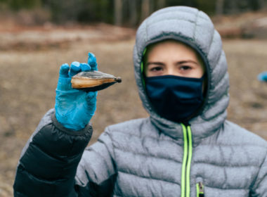
Microplastics are very small plastic pieces less than five millimeters long that originate from a variety of human-made sources, such as plastic water bottles, plastic packaging, textile fibers and cosmetics. They can be harmful to the marine environment and aquatic life. This investigation will focus on microplastics in soft shelled clams. Because soft shelled clams are marine filter feeders, will microplastics be present in clams? If so, how will this effect human health?
Instructions / How to do this investigation
Advanced Preparation
- Choose at least one beach location from which you will collect clam specimens.
- It is very easy to contaminate a sample during collection with plastics from the environment. Do your best under your given circumstances to avoid contaminating your samples with additional plastics. For example:
- Avoid using plastic to collect, store, or filter samples. (If you can’t avoid plastic containers, please don’t let that stop you from participating in the investigation, though.)
- Be careful if you are wearing clothing containing polyester not to let any fibers from your clothes get into the sample, while collecting or back in the lab.
- Keep all collection containers capped until you are ready to use them.
- Read through the field instructions and sample analysis instructions sections below, and be sure to assemble and check all materials ahead of time.
- Choose an adequate container or containers to collect your specimens. Non-plastic is best, but if plastic is all you have, use it.
*Please note that a microplastic is less than or equal to 5 millimeters at its fattest or longest part, whichever is bigger.
Field Instructions
- All samples are to be collected at the same site at or near low tide.
- If you wish to compare over time, feel free to collect samples as frequently as you are able.
- Use a waterproof camera to take a picture of the beach/area from which you will be taking your sample. Take the picture from an angle that captures the spot from which the sample will be taken.
- You may wish to take additional pictures of structures that border the water body.
- Record the following information about your collection site the day of collection.
- Harvest Date
- Time
- Latitude and longitude
- Tide stage
- Direction the beach faces
- Water temperature
- Before sampling, label each container with
- Beach/location name, and a number if using more than one container to collect your clams.
- Example: TownBeach-1, TownBeach-2
- Harvest Date
- Provide a key of any naming system that uses abbreviations.
- Beach/location name, and a number if using more than one container to collect your clams.
- Specimens can be gathered randomly at designated beach locations and placed in the labeled sampling container.
- Specimens can be located by looking for holes approximately the size of an index finger in the mud flats.
- Once a suspected clam hole is found, use a clam rake or your hands to dig about a foot under the mud surface to locate the clam.
- To mitigate the possibility of contaminating the sample with microplastics in sea water.
- Once transported back to the school, clams can be placed in a bag or other container and placed in the freezer for storage.
Sample Analysis Instructions
- Carefully pry open the clam, being sure to not break the shell and retaining all body tissue.
- The body tissue should be removed, weighed in grams and then placed into a labeled 500ml erlenmeyer flask.
- Digestion should be performed using a 40:1 ratio of 30% H2O2.
- This should be 200mL of H2O2 for every 5 grams of clam tissue.
- The solution should then be placed in an incubator for 24 hours at 55°C.
- After 24 hours, the solution should be stored for an additional 48 hours to complete the digestion.
- This mixture should be filtered using the vacuum flask and a .45um filter.
- After the sample has filtered, the mesh can be placed in a petri dish and covered for microscopic analysis.
Decide on amount of analysis to do, and prepare data tables*
- You have three options for sample analysis and data entry, represented by tabs 2-4 of the data entry form on WB . Choose one of these three options.
- Qualitative Data –
- Identify the presence or absence of microplastic fibers and of microplastic fragments/pellets, and list the colors you observe.
- You will complete only one data entry per class on the WB site.
- Type & Color –
- Identify microplastics by type and color, and report the number of each found.
- You will complete one data entry per filter or per gallon on the WB site. If data is entered per filter, add the filter size to the sample name (example: TownBeach-1-50µ).
- Color, Shape & Size –
- For each individual microplastic found, identify and record its color, shape and size.
- There can be multiple entries. Each sample label should include at least the beach name and sample number (example: TownBeach-1).
- Qualitative Data –
Analyze Samples
- Examine a filter using a hand lens or dissecting microscope with low magnification (ex- 4.5x). Increase the magnification as needed (up to 40x). Record your findings on your data sheet.
- If you need a closer look at a particular fragment, you can try to move it to a slide with a drop of water (tricky to do!) and examine it under a compound microscope with higher magnification.
- Some microscopes may fit a petri dish for closer examination under higher power, but you’ll still need to add water.
- Use tweezers and probes to gently poke at fragments on the filter paper or mesh. Most plastic fragments are somewhat flexible and will not break when prodded. Plastic fragments will often bounce or spring when prodded. If a fragment breaks when touched, do not count it as plastic.
- Use The MERI Guide to Microplastics to help you:
- Identify if you are seeing microplastics or something else (see the hints below on p. 6, plus sections E & F on pages 5-10 of the MERI Guide)
- Distinguish fibers (also called filaments) from fragments/pellets and further sort them by shape (see Figure 17 on pages 10-11 of the MERI Guide)
- Enter your data into the appropriate tab on WB (you chose this during step 4 on page 4), and remember:
*Hints for identifying if microplastics are present in sample (The pictures in this document might help you decide if you’re looking at a microplastic or something else.)
- Microplastic fibers are the same thickness all along their length, and tend to have smoother edges then other types of debris.
- Microplastics are often one consistent color throughout the piece.
- Microplastics tend to look less shiny under a microscope than things like pebbles and sand grains.
- Most microplastics are a little flexible and won’t break when prodded with tweezers or a probe. They may even bounce or spring a little.
- If it has cell structures inside it, it’s not a microplastic. However, living things might be attached to a microplastic. If you’re not sure if it’s plastic or living, don’t count it!


Comments
Tell us how your data collection/analysis is going